
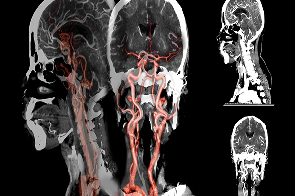
Computed Tomography Angiography, or CTA, is a specialized type of CT scan designed specifically to visualize the body’s arteries and veins. It is a powerful, non-invasive test that can provide highly detailed 3D images of the vascular system, allowing physicians to diagnose a wide range of conditions, from blockages and aneurysms to traumatic
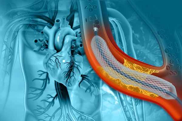
An Introduction to Interventional Radiology: Image-Guided Procedures
Interventional Radiology (IR) is a dynamic and rapidly growing subspecialty of radiology that uses medical imaging to perform minimally invasive procedures. Often described as “surgery through a pinhole,” IR represents a significant shift from traditional open surgery. Interventional radiologists are physicians who use tools like cathet
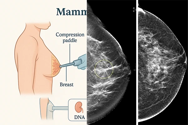
Mammography: The Gold Standard in Breast Imaging
Mammography is a specialized medical imaging technique that uses a low-dose X-ray system to see inside the breasts. For decades, it has been the most effective primary tool for the early detection of breast cancer, capable of identifying abnormalities long before they can be felt. Mammography plays a crucial role in both screening asymptomatic wome
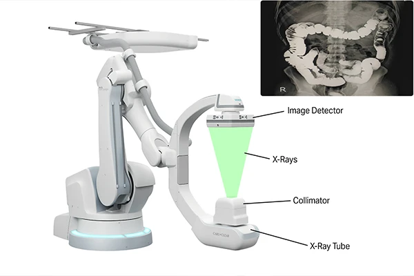
A Guide to Fluoroscopy: Visualizing the Body in Real-Time
If a standard X-ray is like a photograph, then fluoroscopy is like a video. It is a medical imaging technique that uses X-rays to obtain real-time, moving images of the body’s internal structures. This “live X-ray movie” allows physicians and surgeons to observe physiological processes, guide instruments, and perform minimally inv
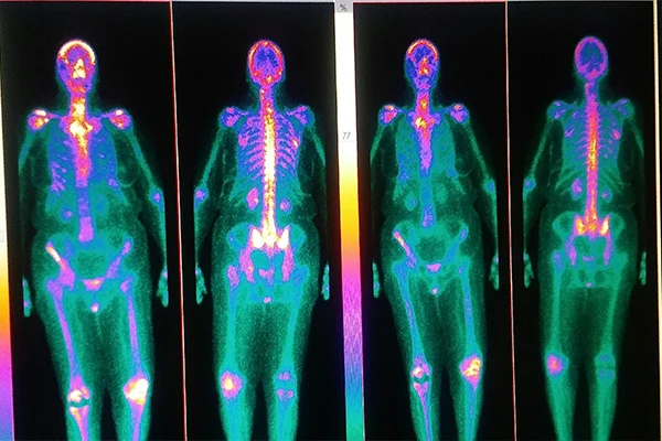
Nuclear Medicine Explained: An Overview of PET and SPECT
Nuclear medicine is a unique branch of medical imaging that provides information about the function of organs and tissues, rather than their structure. While modalities like CT and MRI excel at showing anatomy, nuclear medicine excels at showing physiology. It reveals how the body is working on a cellular and molecular level. The Core Principle: [&
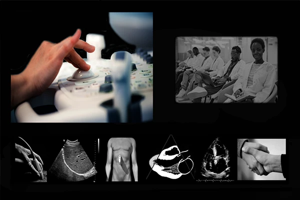
An Introduction to Ultrasound: Seeing with Sound Waves
Ultrasound, also known as sonography, is a widely used medical imaging technique that uses high-frequency sound waves to create real-time images of the body’s internal organs and structures. Unlike X-ray or CT, it does not use any ionizing radiation, making it an extremely safe and versatile tool, especially in sensitive applications like obs
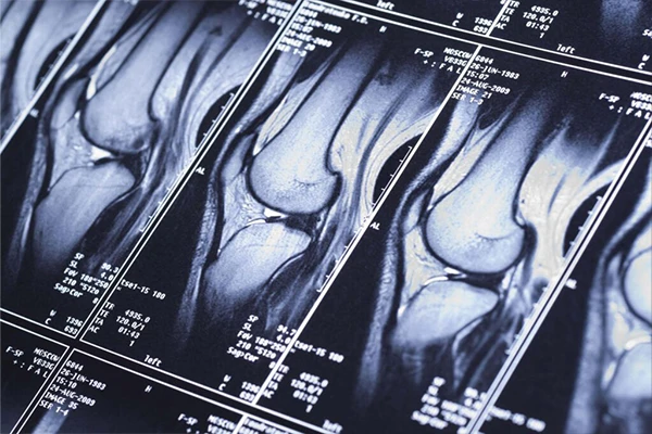
The Principles of Magnetic Resonance Imaging (MRI)
Magnetic Resonance Imaging (MRI) is a sophisticated medical imaging technique that stands apart from X-ray and CT because it does not use ionizing radiation. Instead, it uses a powerful magnetic field, radio waves, and a computer to create exquisitely detailed images of the body’s internal structures. Its greatest strength is its unparalleled
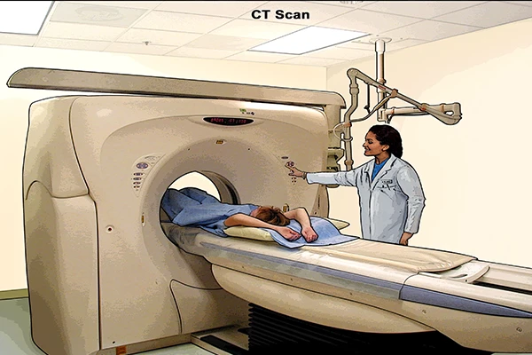
An Introduction to Computed Tomography (CT): Seeing the Body in Slices
While a conventional X-ray provides a flat, two-dimensional view of the body, Computed Tomography (CT) represents a revolutionary leap forward. By combining X-ray technology with powerful computers, CT scanners produce detailed, cross-sectional images—or “slices”—of the body, allowing us to see inside with unprecedented clarity. It elim
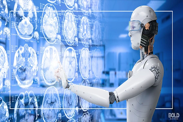
The Future of AI in Radiology: What to Expect in the Next 5 Years
The first wave of Artificial Intelligence in radiology has focused on “seeing”—detecting and highlighting abnormalities. The next wave will focus on “understanding” and “predicting.” As AI models become more sophisticated and integrated into clinical workflows, we can expect to see them take on more complex tasks
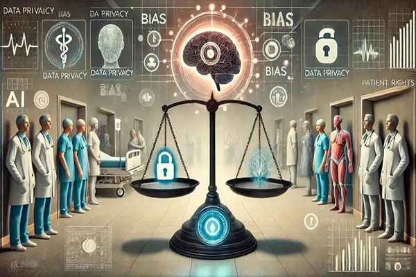
The Ethical Challenges and Biases of AI in Medical Imaging
While Artificial Intelligence holds immense promise for improving medical imaging, its development and implementation come with a host of complex ethical challenges. As we integrate these powerful tools into clinical practice, it is crucial that developers, clinicians, and regulators carefully consider the potential pitfalls to ensure that AI is us