
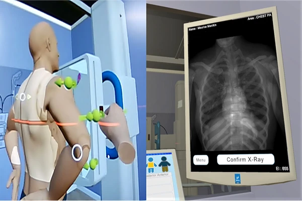
Acquiring a high-quality radiograph is a skill that depends on three factors: setting the correct exposure technique, providing clear instructions to the patient, and, most importantly, precise patient positioning. Proper positioning ensures that the correct anatomy is visualized without distortion or superimposition, which is essential for an accu

The ALARA Principle: A Practical Guide to Radiation Safety
In any field that uses ionizing radiation, from nuclear power to medical imaging, safety is the number one priority. The guiding philosophy for radiation safety in radiology is the **ALARA** principle. ALARA is an acronym that stands for “As Low As Reasonably Achievable,” and it is the cornerstone of every radiation protection program.
DICOM vs. NIfTI: Understanding the Key Differences for Neuroimaging
While DICOM is the undisputed standard for clinical medical imaging, anyone entering the world of academic neuroimaging research will quickly encounter another format: NIfTI. Though both are used to store brain scans, they were designed for very different purposes. Understanding their core differences is key to working effectively in both clinical
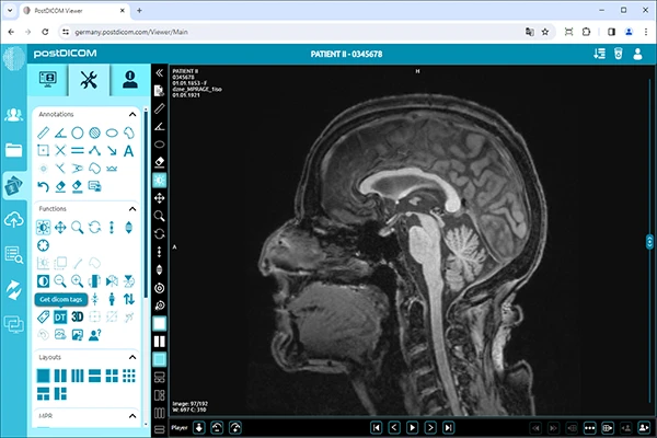
DICOM Tags Explained: A Deep Dive into Medical Image Metadata
A DICOM file is far more than just a picture. It’s a comprehensive data object that bundles the image pixels with an extensive set of metadata. This metadata, organized into “DICOM tags,” is what gives an image its clinical context, ensuring it is never separated from critical patient and study information. Understanding these tag
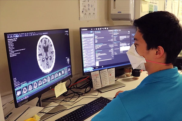
An Introduction to PACS and RIS: The IT Backbone of Radiology
Behind every modern radiology department is a sophisticated network of software that manages everything from patient scheduling to image storage. The two most critical components of this digital ecosystem are the PACS and the RIS. While they work together seamlessly, they have very different functions. Understanding the role of each is essential fo
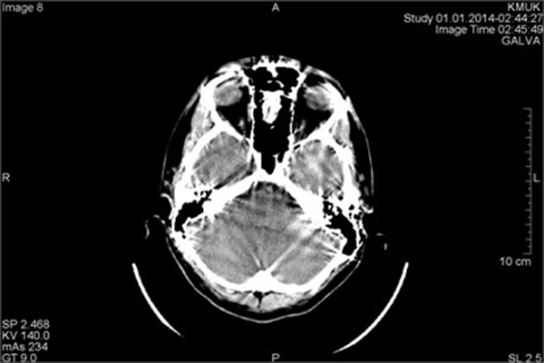
Common Imaging Artifacts (CT & MRI) and How to Troubleshoot Them
In a perfect world, every medical image would be a flawless representation of the patient’s anatomy. In reality, a variety of factors can degrade image quality, creating “artifacts”—features seen in an image that are not present in the actual object. For a technologist, recognizing, understanding, and preventing artifacts is a cri
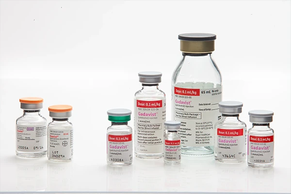
The Role of Contrast Agents in Radiology: A Practical Guide
In medical imaging, contrast agents—often called “contrast media” or “dyes”—are special substances used to improve the visibility of internal body structures. Many soft tissues have similar densities and can be difficult to differentiate on a standard scan. Contrast agents temporarily change the way imaging systems interact
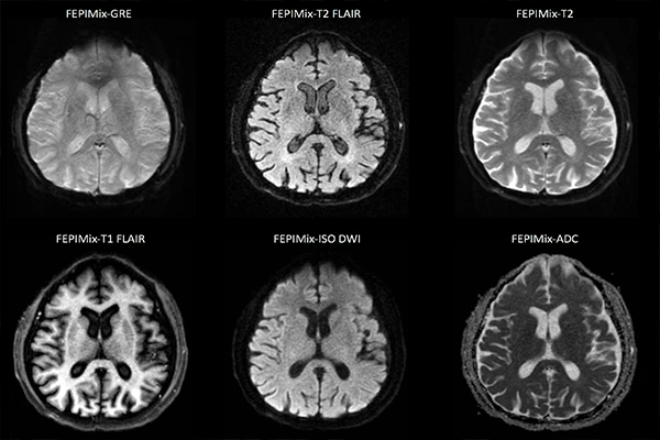
Understanding MRI Sequences: A Guide to T1, T2, FLAIR, and DWI
The power of MRI lies in its versatility. By adjusting the timing and parameters of the radiofrequency (RF) pulses and signal detection, technologists can generate a variety of “image weightings” or sequences. Each sequence is designed to highlight specific tissue characteristics, providing different pieces of the diagnostic puzzle. A s
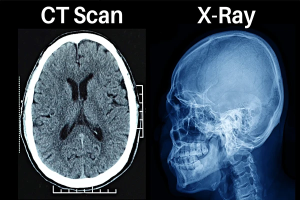
X-ray vs. CT Scan: What’s the Difference?
Both X-rays and CT (Computed Tomography) scans are common medical imaging tests that use X-ray radiation to see inside the body. While they are based on the same fundamental technology, they produce very different types of images and are used for different reasons. Think of a standard X-ray as a simple photograph and a CT […]
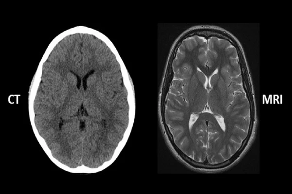
CT vs. MRI: A Detailed Comparison for Technologists
Computed Tomography (CT) and Magnetic Resonance Imaging (MRI) are two of the most powerful diagnostic tools in modern radiology. While both produce detailed, cross-sectional images of the body, they are fundamentally different technologies with distinct strengths, weaknesses, and clinical applications. Understanding when and why to use one over the